Scientists Broaden Near-Infrared Fluorescence Imaging’s Unexplored Window Scope
Date:07-08-2018 | 【Print】 【close】
A research team led by Prof. CAI Lintao at the Shenzhen Institutes of Advanced Technology (SIAT) of the Chinese Academy of Sciences extended near-infrared fluorescence imaging window ranges, which has markedly enhanced signal-to-background ratio because autofluorescence, scattering and light absorption by biological tissues and water are weaker at longer wavelengths.
Their related work was published in Theranostics with the title “Near-infrared fluorescence imaging in the largely unexplored window of 900–1,000 nm”.
Near-infrared (NIR) fluorescence imaging is a powerful technique for visualizing deep-tissue structures, like blood vessels, lymph nodes and detecting tumors.
Currently, the scientific community posits that there are two biologically transparent windows where NIR fluorescence imaging can be performed: one called NIR-Ia (700–900 nm), the other called NIR-II (1,000–1,700 nm) window. Most people have been avoiding the NIR-Ib (900–1,000 nm) window due to the lack of suitable fluorophores and the presence of a water overtone absorbance peak.
In this study, they found that six heptamethine dyes had distinct emission peaks in both the NIR-Ia and NIR-Ib window, and they demonstrated that NIR-Ib fluorescence imaging was clearer, sharper and more penetrative than NIR-Ia fluorescence imaging. The technique could yield images of lymph nodes, subcutaneous tumors, brain tumors and — for the very first time — leaf veins and early infected anthracnose of plant leaves.
To gain molecular insights into how NIR-Ib fluorescence imaging works, they performed linear spectral unmixing analysis — typically used in remote sensing but has never used for our purpose — to show that autofluorescence and scattering were reduced in the NIR-Ib window.
"This work not only contests a widespread assumption in NIR fluorescence imaging but also have important implications in botany, ecology and medical research” said Prof. CAI.
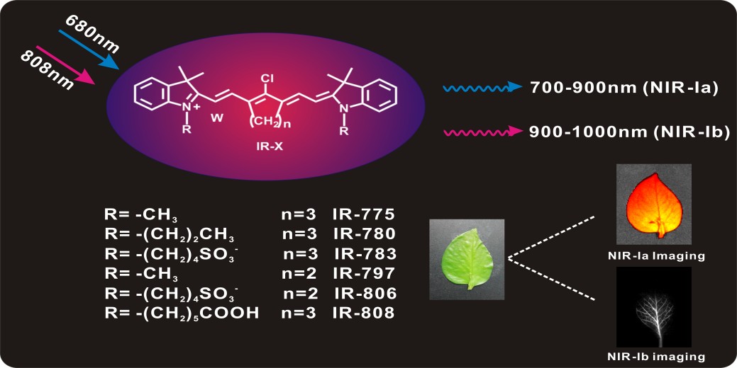
Heptamethine dyes had distinct emission peaks in both the NIR-Ia and NIR-Ib window, and the technique applied for plant vein imaging (Image by Prof. CAI)