
Dedicated 36-Channel Receive Array Breaks Through the Limitation of Existing MRI for Fetal
Date:06-06-2018 | 【Print】 【close】
Magnetic resonance imaging (MRI) has been an important tool for defining fetal anatomy and pathology non-invasively due to its non-ionizing radiation feature, superior soft tissue contrast, and the capability of a large field-of-view (FOV) scan to image the entire body of the fetus, especially for the diagnosis of neurodevelopmental disorders and cardiovascular diseases.
Due to a lack of fetal imaging coils, the standard commercial abdominal coil is often used for fetal imaging, the performance of which is limited by its insufficient coverage, element number and signal-to-noise ratio (SNR).
A research team led by Prof. ZHENG Hairong at the Shenzhen Institutes of Advanced Technology (SIAT) of the Chinese Academy of Sciences, developed a dedicated 36-channel coil array for fetal imaging at 3 T, with high density coil elements, wide coverage and flexibility to conform to the varied anatomy of pregnant patients.
Compared to a commercial abdominal coil array, the proposed 36-channel fetal array provides not only SNR improvements, up to 40%, but also an augmented imaging speed. Clear fetal brain (twice as fast) and heart movie (4 times faster) imaging were successfully performed with the dedicated 36-channel coil array.
This work named “A Dedicated 36-channel Receive Array for Fetal MRI at 3 T”, was published in IEEE Transactions on Medical Imaging.
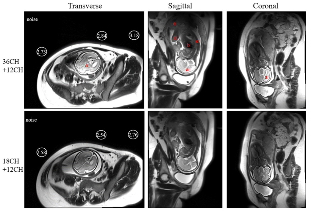
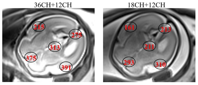
Figure 1. Fetal images in the transverse, sagittal and coronal planes acquired by using the proposed 36-channel coil and the commercial abdominal coil both combined with a 12-channel spine coil. (Image by ZHENG Hairong)
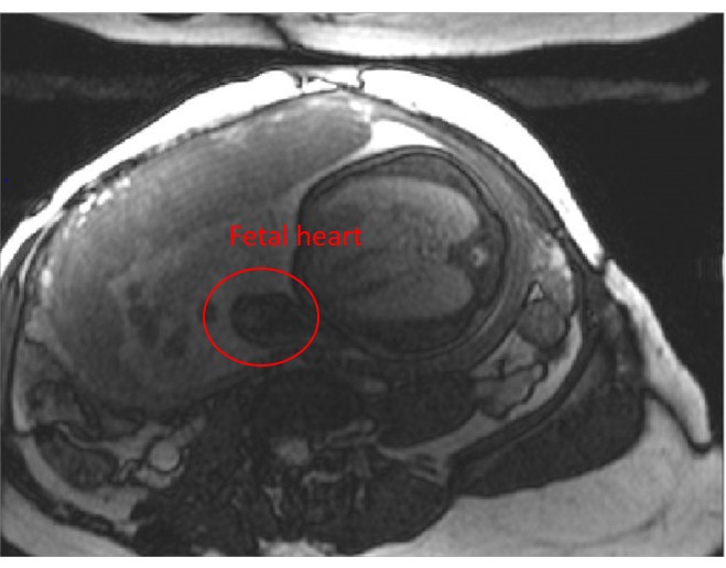
Figure 2. Fetal heart image in the transverse planes acquired by using the proposed 36-channel coil combined with a 12-channel spine coil. (Image by ZHENG Hairong)