
Ring Artifacts Correction for Computed Tomography Image Using Unsupervised Contrastive Learning
Date:24-10-2023 | 【Print】 【close】
Researchers led by Associate Professor LIANG Xiaokun from the Shenzhen Institute of Advanced Technology (SIAT) of the Chinese Academy of Science (CAS), along with their collaborators have proposed a novel ring artifact removal algorithm based on double contrast learning (DCLGAN) and an image style transformation network.
The study was published in Physics in Medicine & Biology on Oct. 4.
Computed tomography (CT) is an imaging technique that generates three-dimensional images from two-dimensional projection data. CT is a widely employed imaging technology for disease detection.
However, CT images often suffer from ring artifacts, which may result from hardware defects and other factors. These artifacts compromise image quality and impede diagnosis.
In this study, employing DCLGAN to eliminate strip artifacts in the polar coordinate system. Subsequently, applying post-processing techniques to extract ring artifacts from the image, with the aim to remove them.
In detail, the proposed method involved simulating ring artifacts on real CT data to generate the uncorrected CT (uCT) data and transformed them into strip artifacts.
Subsequently, the DCLGAN synthetic network was applied in the polar coordinate system to remove the strip artifacts and generate a synthetic CT (sCT). Then, researchers compared the uCT and sCT images to obtain a residual image, which was then filtered to extract the strip artifacts. An inverse polar transformation was performed to obtain the ring artifacts, which were subtracted from the original CT image to produce a corrected image.
Researchers used real CT data to validate the effectiveness of the proposed approach. Results demonstrated that the corrected CT images showed a reduction in mean absolute error by 12.36 Hounsfield units (HU), a decrease in root mean square error by 18.94 HU, an increase in peak signal-to-noise ratio by 3.53 decibels (dB), and an improvement in structural similarity index by 9.24%.
Furthermore, researchers conducted a comprehensive evaluation of the proposed method, assessing its performance not only on real CT data but also on synthetic data and patient?s brain cone beam computed tomography (CBCT) data that were not part of the training dataset. External testing on synthetic data and CBCT images of the patient's brain further demonstrated the method's effectiveness in effectively reducing ring artifacts in the CT images.
"Our method demonstrates excellent performance across various datasets, highlighting its strong adaptability. This approach notably suppressed ring artifacts in CT images while preserving image structure and fine details. It proves to be a practical and appealing solution for applications in CT-guided radiotherapy and disease diagnosis." said Prof. LIANG.
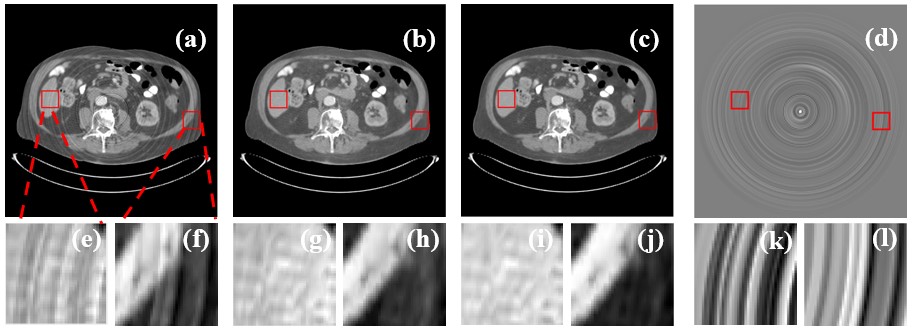
Results of the proposed method on CT images of the patient’s abdomen. (Image by SIAT)
Media Contact:
ZHANG Xiaomin
Email:xm.zhang@siat.ac.cn