A Capsule X-Ray Dosimeter for Real-Time Radiotherapy Monitoring
Date:13-04-2023 | 【Print】 【close】
In radiotherapy, precision in targeting tumor tissue while minimizing damage to healthy tissue is crucial. Monitoring the dose of radiation delivered and absorbed in real-time, particularly in the gastrointestinal tract, poses significant difficulty. Additionally, existing methods used for monitoring biochemical indicators such as pH and temperature are inadequate for comprehensive evaluation of radiotherapy.
To address this challenge, a joint research team from the Shenzhen Institute of Advanced Technology (SIAT) of the Chinese Academy of Sciences, the National University of Singapore (NUS), and Tsinghua University has developed a capsule-shaped swallowable X-ray dosimeter, which can estimate radiation dose based on radioluminescence and temperature using a neural network-based regression model.
The researchers found that the dosimeter was approximately five times more accurate than standard methods for dose determination.
The study was published in Nature Biomedical Engineering on April 13.
In vivo clinical dosimeters such as metal-oxide-semiconductor field-effect transistors, thermoluminescence sensors, and optically excited films are commonly placed directly on or near the patient’s skin to estimate the dose absorbed in the target area.
Although in vivo dosimetry with electronic portal imaging devices has been explored for treatment verification, it can be expensive and subject to photon attenuation that can alter the dose to the patient.
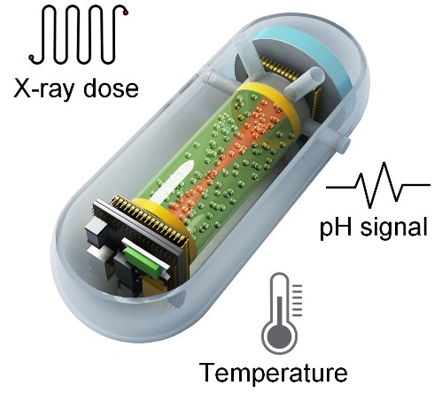
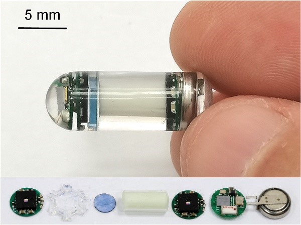
The capsule dosimeter is composed of a flexible optical fiber encapsulated with X-ray persistent nanoscintillators, a polyaniline film, and a wireless miniaturized luminescence readout system.
With the capability of measuring pH and temperature, it can evaluate the absorbed dose during radiotherapy for gastric cancer and can be used to monitor treatment for different malignancies with further optimization of the capsule’s size.
“In the future, this capsule could be placed in the rectum for prostate cancer brachytherapy or in the upper nasal cavity for real-time measurement of the absorbed dose in nasopharyngeal carcinoma minimizing radiation damage to surrounding structures,” said Prof. SHENG Zonghai, one of the corresponding authors.

Figure 1. Functional model of capsule. (Image by SIAT)

Figure 2. Separate component model of capsule. (Image by SIAT)

Figure 3. Photo image of capsule. (Image by SIAT)
Media Contact:
ZHANG Xiaomin
Email:xm.zhang@siat.ac.cn