Deep Multi-Magnification Similarity Learning for Histopathological Diagnosis
Date:07-02-2023 | 【Print】 【close】
Precise classification of histopathological images is the ultimate objective of the image analysis.
In actual clinical applications, pathologists usually combine information at different magnifications, that is, from subnuclear O (0.1 μ m) to cells (≈ O (10 μ m)) and intercellular (≈ O (100 μ m)), to other larger tissues (≈ O (1mm)) for diagnosis.
Magnification-based learning networks, which usually combine information at different magnifications, have attracted considerable attention for their ability to improve performance in histopathological classification.
However, fusion of various images has not been studied in depth.
Recently, a research team led by Dr. QIN Wenjian from the Shenzhen Institute of Advanced Technology (SIAT) of the Chinese Academy of Sciences, together with the Prof. LUO Weiren team from Shenzhen Third People's Hospital and Prof. Nazar Mustafa Zaki team from United Arab Emirates University, UAE, proposed a novel deep multi-magnification similarity learning (DSML) approach to improve performance in?histopathological classification.
The work was published in the IEEE Journal of Biomedical and Health Informatics on January 16.
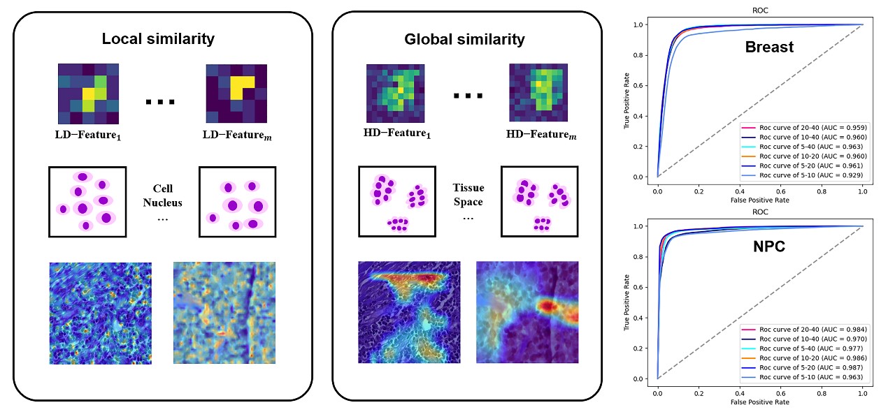
The DSML focuses on tumor histopathological diagnosis by deep learning on digital pathology, which can be useful for the interpretation of multi-magnification learning framework and easy to visualize feature representation from low-dimension (e.g., cell-level) to high-dimension (e.g., tissue-level).
"It can overcome the difficulty of understanding cross-magnification information propagation," said Dr. QIN. "It uses a similarity cross-entropy loss function designation to learn the similarity of the information among cross-magnifications simultaneously."
The researchers designed different network backbones and magnification combinations to verify the effectiveness of DMSL on a clinical nasopharyngeal carcinoma and a public breast cancer BCSS2021 dataset.
They also investigated its ability to interpret. The results showed that it performed better in classification with a higher value of area under curve, accuracy, and F-score than other comparable methods.