Albumin-Consolidated Dyes: NIR-II Fluorescence Imaging Probes for Brain Imaging
Date:27-06-2022 | 【Print】 【close】
Fluorescence imaging in the second near-infrared window (NIR-II, 1,000-1,700 nm) as an important tool for fundamental and clinical research has been greatly developed in the last decade due to its deep penetration, high sensitivity, and high spatial and temporal resolution. It is an indispensable tool for multi-scale molecular imaging ranging from subcellular to living systems. Fluorescent probes with high brightness and active-targeting ability are of great importance to achieving high-quality images.
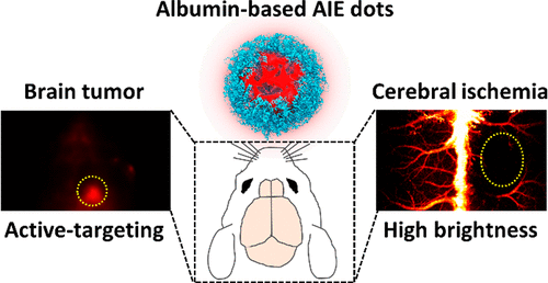
Now, a research team led by Dr. SHENG Zonghai from the Shenzhen Institute of Advanced Technology (SIAT) of the Chinese Academy of Sciences has reported a strategy using endogenous albumin as a carrier. The albumin-based NIR-II fluorescent probe with high fluorescence brightness was constructed and achieved active-targeting NIR-II imaging of brain tumors and cerebrovascular imaging with a high signal-to-background ratio and high resolution.
The study was published in ACS Applied Materials & Interfaces on Jan. 7.
"Endogenous albumin has a specific three-dimensional spatial structure and hydrophobic domain," said Dr. GAO Duyang, the co-author of this study, "resulting in the formation of stable probes through hydrophobic interaction and hydrogen bond."
The interaction can effectively inhibit the intramolecular vibration of dyes and lead to enhanced fluorescence quantum yield. In addition, the albumin-consolidated strategy endows the probe's active tumor-targeting ability since albumin can be endocytosed into tumor cells mediated by the gp60 receptor.
The results showed that the quantum yields of three kinds of albumin-based probes with fluorescence emissions ranging from the visible (400–650 nm) to the NIR-II region were at least 10% higher, and the tumor-targeting efficiency was ~25% higher, compared with those of nanoprobes constructed by the exogenous polymer.
For further in vivo imaging, orthotopic glioma can be observed after intravenous injection of the albumin-based probes, indicating that the albumin-based probes could be accumulated in glioma through the blood-brain barrier (BBB). In contrast, Traditional probes modified by DSPE-PEG-2000 are difficult to achieve glioma imaging by the enhanced permeability and retention (EPR) effect. In addition, albumin-based probes have achieved cerebrovascular imaging with a high signal-to-background ratio (SBR, ~90) and high resolution (~70 μm) in mouse models due to their excellent properties.
"This study has provided a good tool for imaging-guided surgery of brain tumors and cerebral ischemia,” said Dr. SHENG, the corresponding authors of this study “which will improve surgical efficacy to prevent recurrence and side effects."

Albumin-based NIR-II fluorescent probe with high fluorescence brightness was constructed. (Image by Dr. SHENG)
Media Contact:
ZHANG Xiaomin
Email:xm.zhang@siat.ac.cn