Whole Brain Mapping of Primates Enters Micron Era
Date:27-07-2021 | 【Print】 【close】
The research team of Prof. Guo-Qiang BI and Prof. Pak-Ming Lau from University of Science and Technology of China (USTC) and Shenzhen Institute of Advanced Technology (SIAT) of the Chinese Academy of Sciences, along with domestic and international collaborators, realized three-dimensional mapping of the entire brain of the macaque monkey at micron resolution, based on an updated version of their recently developed high-throughput three-dimensional fluorescence imaging technique VISoR, and an efficient pipeline SMART. This work is published in Nature Biotechnology on July 26th.
Our brain comprises nearly a hundred billion nerve cells with delicate and complex connections among them. To fully understand how the brain functions, it is essential to have a high-resolution map of how the nerve cells are organized and connected within the brain. At present, it usually takes days to complete three-dimensional imaging of the whole brain of a mouse at micron resolution using the current state-of-the-art techniques.
However, high-resolution brain mapping for nonhuman primates such as the rhesus monkey, although highly desired given their roles in modeling human diseases, has been hindered by a major technical challenge. The brain of a rhesus monkey is too big, more than 200 times larger in volume than that of a mouse.
To overcome this challenge, the team developed a high-throughput three-dimensional fluorescence imaging technique, Volumetric Imaging with Synchronous on-the-fly-scan and Readout, VISoR. Compared to commonly used 3D optical imaging techniques, VISoR eliminates the time loss caused by moving and pausing while switching fields of view to obtain 2D image series, allowing for imaging without blur when the sample is in continuous motion. In this way, VISoR achieves extremely high speed, dozens of times faster than other 3D imaging methods for large tissue samples.
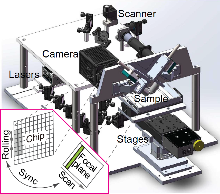
Schematic of VISoR2 (Image by XU Fang et al.)
In addition to the challenge of imaging throughput, difficulty of imaging monkey brains also arises from the complicated cortical folding structures and low tissue transparency. The team began with embedding of the isolated brain followed by sectioning. The tissue sections were then permeated and refractive index matched by using a primate-optimized uniformly clearing (PuClear) solution. With their improved VISoR2 system, they finished imaging of a whole macaque monkey brain.
"Brain connectome at the mesoscopic level is important but so far limited to rodents. This work demonstrates a powerful method that enables researchers to dissect mesoscopic connectome of monkeys at one micron resolution, in four days. It represents a tour de force in this rapidly moving field." Prof. Xiaojing Wang from the New York University commented.
The total volume of raw image data acquired from two macaque brains exceeded 1 PB. The team further developed efficient algorithms and software to realize automated 3D imaging reconstruction and semi-automated long-distance tracking of individual neuronal axon fibers. Their initial observations revealed previously unknown characteristics of axonal fiber projection and surprising patterns of fiber turning and routing in the cortical folds.

Axon projection and tracing of thalamic neurons in rhesus monkeys (Image by XU Fang et al.)
Prof. David C. Van Essen from Washington University in St Louis describes this work as a "technical tour de force that marks a stunning advance in our ability to map long-distance connectivity accurately and efficiently throughout the entire brain” of the macaque monkey. Besides the technical achievement, their “exciting discovery may have profound implications for understanding brain morphogenesis and the principle of 'wiring length minimization'," he comments.
Besides brain mapping, the application of VISoR may be extended to imaging of other tissues and organs, including samples from clinical pathology. It is anticipated that by combining the obtained huge imaging data with AI analysis, this technique may eventually help us understand the fine 3D structure of the brain and body as well as how they change in various disease conditions, and thus facilitating medical diagnostics and drug developments.
"It is hoped that this technology will be further improved on the application level, and could be promoted and applied on a large scale, and make an important contribution to the mapping and understanding of primates and even the human brain." Prof. Shumin Duan from Zhejiang University outlooked.
Written by JIANG Pengcen, USTC News Center
Media Contact:
SUN Lujia
Email: lj.sun @siat.ac.cn
Download the attachment: