
Scientists Develop Biomimetic Nanoprobes to Image Brain Tumors
Date:10-08-2020 | 【Print】 【close】
Glioma is the most malignant tumor of the central nervous system. The diagnosis and treatment of glioma are great challenges due to the presence of the blood brain barrier (BBB) and the complex brain tumor microenvironment.
Researchers from the Shenzhen Institutes of Advanced Technology (SIAT) of the Chinese Academy of Sciences collaboration with the National University of Singapore developed biomimetic nanoprobes for targeted imaging of brain tumors.
The study was published in Advanced Functional Materials.
Researchers firstly prepared the biomimetic nanoprobes mainly by composing ferric oxide and fluorescent dye, and the surface of nanoprobes was coated by bioorthogonally labeled brain tumor cell membrane for crossing the BBB and targeting brain tumor cells.
As a diffuse tumor, glioma normally could not be excised en‐bloc with a large margin, which places stringent demands for selective localization and imaging of tumor margin at a microscopic level. Researchers and surgeons used the fluorescence, photoacoustic and magnetic resonance imaging for tumor margin identification, as well as real‐time image‐guided resection.
Then through cellular uptake and mice model experiments, the combination of brain tumor cell membrane-based biomimetic technology and peptide-based active-targeting features improved the efficacy of diagnosis of brain tumors greatly.
This study provided some insights on theranostic targeting of brain tumor cells from the perspective of physiochemistry, gave edging closer to a thorough understanding of brain tumor targeting mechanisms.
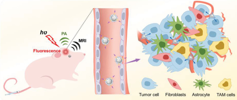
Schematic illustration of the design of functionalized nanocomposites for BBB penetration and navigation through brain tumor microenvironment (Image by SIAT)
Media Contact:
ZHANG Xiaomin
Email: xm.zhang@siat.ac.cn