
MRI Study Reveals Arterial Culprit Plaque Characteristics
Date:01-07-2020 | 【Print】 【close】
The degree of cerebral infarction (single or multiple infarcts) is a feasible imaging marker to predict future stroke recurrence in patients with ischemic stroke. However, the features of arterial vessel wall lesions and culprit plaques ultimately leading to different degree of cerebral infarction still remains unclear.
High resolution magnetic resonance vessel wall imaging (HR-MRVWI) provides unique advantages in detecting and directly visualizing arterial vessel wall lesions and plaques, which has been demonstrated to be superior to arterial lumen imaging.
Researchers from the Shenzhen Institutes of Advanced Technology (SIAT) of the Chinese Academy of Sciences developed a head-neck combined HR-MRVWI technique, which could potentially provide remarkable suppression of cerebrospinal fluid signals, enhanced T1 (Longitudinal relaxation) contrast weighting, and most importantly, the whole-brain and neck spatial coverage.
The study was published in Magnetic resonance imaging.
By using the head-neck combined HR-MRVWI technique, researchers compared the characteristics of culprit plaques among different degree or types of infarction.
This specific head-neck coverage was capable of depicting arterial vessel wall lesions of multiple vascular beds from common carotid arteries to distal segments of intracranial arteries, which was for comprehensive evaluation of ischemic stroke etiology and recurrence risk.
The experiment results showed that more culprit plaques per patient were found in multiple-infarction group than single-infarction group, as well as in non-perforating artery infarction (PAI) group than PAI group.
For patients with large artery atherosclerosis induced ischemic stroke, those with multiple infarcts had more severe arterial stenosis and more prominent plaque enhancement than those with single infarct. For patients with anterior circulation ischemic stroke, those with PAI showed lower arterial stenosis degree than non-PAI.
This work confirmed that there’s positive correlation between stenosis grade/plaque vulnerability and multiple infarcts occurrence, that could potentially increase the risk of stroke recurrence.
Besides, even a plaque with not obvious stenosis on angiography could contribute to the occurrence of PAI, that is to say, the degree of plaque enhancement might be a feasible imaging marker to predict the degree of infarction, which would be useful for supporting therapeutic decision-making and risk assessment.
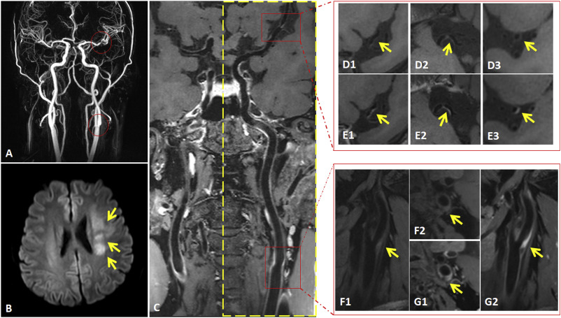
An example of multiple infarcts based on (diffusion-weighted imaging) DWI image with culprit plaques detected on both intracranial middle cerebral artery and extracranial carotid artery on HR-MRVWI image. (Image by SIAT)
ZHANG Xiaomin
Email: xm.zhang@siat.ac.cn