
Porcine Study Shows New Finger Size Ultrasound Capsule Endoscopy Capture Effective High-Resolution Image
Date:10-03-2020 | 【Print】 【close】
A research team led by Prof. QIU Weibao from Shenzhen Institutes of Advanced Technology, Chinese Academy of Sciences introduced a novel μUSCE (micro-ultrasound capsule endoscopy), uses a capsule equipped with μUS (micro-ultrasound) transducers, capable of imaging below the GI wall surface, offering thereby a complementary sensing technique to optical imaging capsule endoscopy.
The structure of the μUSCE device, shown in Figure1, is consists of a micromotor, a transducer holder and an ultrasound transducer, encased in a biocompatible shell with an acoustically transparent window.
The USCE shell of diameter 10 mm and length 30 mm was made from biocompatible poly (methyl methacrylate) (PMMA) (Z = 3.2 MRayl). The μUS transducer was fabricated from a piezocrystal LiNbO3 to operate at a resonant frequency ?30 MHz, with a press-focused focal distance of 8 mm and the diameter of 3 mm.
The transducer is placed in a PMMA holder that aligns it with both the central longitudinal axis of the capsule and the acoustic window.
The USCE is connected to external instrumentation that drives the motor and controls ultrasound transduction via a Teflon tube of outer diameter 1.7 mm and inner diameter 1.1 mm.
All components and bonding were achieved with medical grade epoxy (Epoxy Technologies, Billerica, MA, USA).
An imaging platform was developed specifically for the evaluation of the proposed μUSCE device. The control of the motor back and forth rotation was realized through two analogue switch channels to control the supply of positive and negative power inputs.
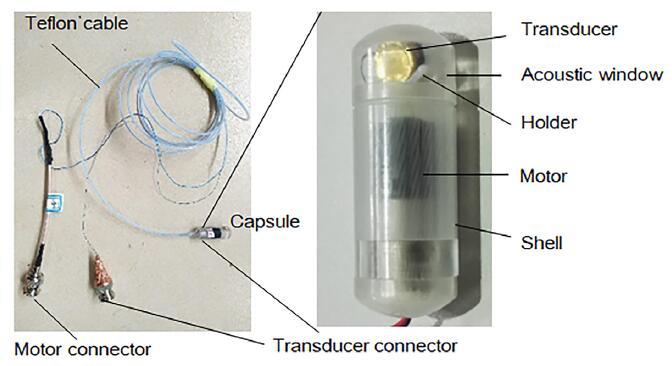
Figure 1. The final μUSCE device (Image by Prof. QIU Weibao)
To demonstrate the effectiveness of the device, in vivo studies were performed in the female Landrace pigs, because they have GI tract anatomic and physiologic similarities with humans.
B-scan data of the small intestine and the oesophagus were successfully recorded during the in vivo experiments, the examples of post-processed images, were shown in Figure 2.
It demonstrated that micro-ultrasound with a mechanical rotation scheme is feasible for assessing the GI tract by providing high-resolution ultrasound images.
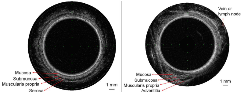
Figure 2. A post-processed cross-sectional ultrasound scan transmural image of a pig small intestine (left) and esophagus (right). (Image by Prof. QIU Weibao)
The proposed scheme in this study paves the way for the implementation of capsule ultrasound devices, and demonstrated the possibility of this capsule by the high-resolution, cross-sectional in vivo imaging.
"This video capsule endoscopes conformed to commercial dimensions and found to be acceptable for clinical use. But the length and shell of the capsule require more engineering work and careful design for further development of ultrasound capsule endoscopes.” said Dr. HUANG Yaocai.
The results were published in Ultrasound in Medicine & Biology.
Media Contact:
ZHANG Xiaomin
Email: xm.zhang@siat.ac.cn
Tel: 86-755-86585299