
Researchers developed 3D super-resolution imaging at unprecedented depth
Date:19-07-2017 | 【Print】 【close】
Optical imaging is a powerful tool in the biomedical sciences, allowing direct inspection and biological processes analysis. Such imaging becomes even more powerful when implemented in three dimensions (3D), and can be further augmented if combined with super-resolution techniques that uncover fine details otherwise obscured by diffraction. Unfortunately, neither 3D nor super-resolution- techniques reach their full potential in thick samples, as optical aberrations (distortions due to sample induced inhomogeneity in refractive index) severely degrade resolution and signal.
ZHENG Wei, the Principle Investigator of Shenzhen Institutes of Advanced Technology, Chinese Academy of Sciences, together with Hari Shroff, the Professor in National Institute of Biomedical Imaging and Bioengineering, developed a new two-photon excitation Instant Structured Illumination Microscope (2P-ISIM) that incorporates rapid adaptive optics (AO) correction enabling 3D and super-resolution imaging achieve unprecedented depth (> 250μm from the coverslip) . This instrument enables ‘real time’ super-resolution imaging. The higher resolution information derived from the excitation and emission point spread functions is directly ‘written’ into the image without the post-processing required in traditional SIM.Adaptive correction is also convenient and fast, since the two photon induced fluorescence functions as a guide star that allows direct wavefront sensing and subsequent aberration correction with a deformable mirror.
This instrument was tested by imaging more than ten different samples, including bead phantoms, cells in gels, Drosophila brain lobe, excised mouse tissue, and live zebrafish and nematode larvae. In all these cases, AO is crucial for both maintaining super-resolution at depth (up to 176±10 nm laterally and 729±39 nm axially, more than doubling resolution relative to conventional 2P imaging), and in boosting signal (up to 40-fold) that would otherwise be lost due to aberrations.
The paper named "Adaptive optics improves resolution and signal in multiphoton super-resolution imaging" is published online on Nature Methods on June 19, 2017. (DOI: 10.1038/nmeth.4337). This work is supported by the National Key Basic Research (973) Program of China, the National Natural Science Foundation of China and the Shenzhen Science and Technology Innovation Committee grant.
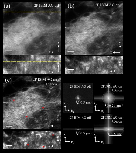
Fig 1. Adaptive optics correction improves spatial resolution of 2P-ISIM in Drosophila larval brain sample. Lateral (top) and axial (bottom) 2P ISIM images of Alexa Fluor 488 phalloidin labeled actin in fixed larval brain lobe, shown without (a) and with (b) adaptive correction, and after subsequent deconvolution (c). The resolution was quantified in MTFs in (d). Reprint from Nature Methods.
Contact ZHENG Wei
Email: zhengwei@siat.ac.cn
Shenzhen Institutes of Advanced Technology